
Functional Units
of the Kidney
# Histologicaly, kidney consists of:
* Cortex: the outer part # granular cuz of glomeruli.
* Medulla : the inner part # striated cuz of tubules.
* Kidney lobule ----> papillae ----> minor calyx ----> major calyx ----> renal pelvis ----> ureter.
Stroma :
1- Capsule
2- Reticular fibers
3- Collagenous and elastic fibers
Parynchema: uroniferous tubules:
1- nephron
2-collecting tubule
Stroma :
1- Capsule
2- Reticular fibers
3- Collagenous and elastic fibers
Parynchema: uroniferous tubules:
1- nephron
2-collecting tubule


The nephron cosists of:
1- Renal corpuscle: # glomerulus # Bowman's capsule.
2- proximal tubule
3- loop of Henle's
4- distal tubule
1- Renal corpuscle: # glomerulus # Bowman's capsule.
2- proximal tubule
3- loop of Henle's
4- distal tubule
# Nephron is functional unit of the kidney.

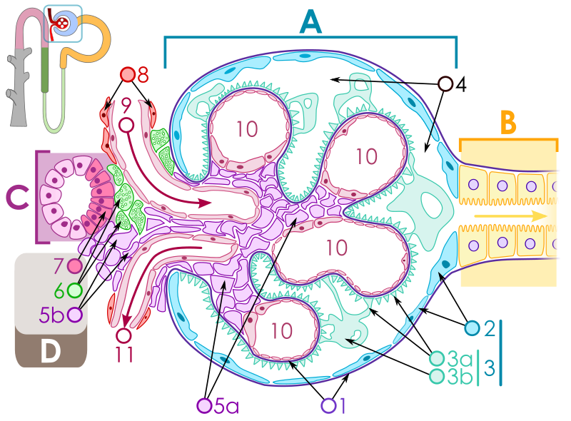
A – Renal corpuscle
B – Proximal tubule C – Distal convoluted tubule D – Juxtaglomerular apparatus 1. Basement membrane (Basal lamina) 2. Bowman's capsule – parietal layer 3. Bowman's capsule – visceral layer 3a. Pedicels (Foot processes from podocytes) 3b. Podocyte |
4. Bowman's space (urinary space)
5a. Mesangium – Intraglomerular cell 5b. Mesangium – Extraglomerular cell 6. Granular cells (Juxtaglomerular cells) 7. Macula densa 8. Myocytes (smooth muscle) 9. Afferent arteriole 10. Glomerulus Capillaries 11. Efferent arteriole |
# Bowman's Capsule: consists of 2 layers:
1- Parietal layer: simble squamous epi. 2
2- Visceral layer: Podocytes 3
# Mesangial cells : # macrophages # supporting cells # part of juxtaglomerular apparatus.
# Proximal tubules : # cubical # wider in diameter # narrower lumen # cells with microvilli # section shows 3-5 cells # cells are indistinct.
# Distal tubules : # cubical # wider in lumen # narrower diameter # cells without microvilli # section shows 5-8 cells # cells are distinct.
# Juxtaglomerular apparatus : # consists of:
1- JG cells: modified SM cells of afferent arteriole.
2- Macula densa: modified cells of distal tubule.
3- Polar cushion: extrglomerular mesangeal cells.
# function of JGA : secretion of Renin
Angiotensinogen ---> angiotensin I ---> angiotensin II


Glomerular Filtration Barrier:
1- Capillary endothelium: fenestrated epi.
2- Fused basement membrane: of 3 layers, the middle is dark
3- Fitration slits diaphragm: between minor - foot - processes of podocytes # covered by diaphragm.


Nephron structure in details:
ليست هناك تعليقات:
إرسال تعليق