about

I'M JUST A MEDICAL STUDENT, TRY TO MAKE THINGS EASY

الجمعة، 1 يونيو 2012

Systemic Lupus Erythematosus

Systemic Lupus Erythematosus 

Autoimmune disease characterized by autoimmune antibodies espesially antinuclear antibodies ANA. 


lupus and Dr. House XD

SYMPTOMS:

  • Chest pain when taking a deep breath
  • Fatigue
  • Fever with no other cause
  • General discomfort, uneasiness, or ill feeling (malaise)
  • Hair loss
  • Mouth sores
  • Sensitivity to sunlight
  • Skin rash -- a "butterfly" rash over the cheeks and bridge of the nose affects about half of people with SLE. The rash gets worse in sunlight. It's called: Malar erythema.
  • Other symptoms depend on what part of the body is affected:
    • Brain and nervous system: headaches, numbness, tingling, seizures, vision problems, personality changes
    • Digestive tract: abdominal pain, nausea, and vomiting
    • Heart: abnormal heart rhythms (arrhythmias)
    • Lung: coughing up blood and difficulty breathing
    • Skin: patchy skin color, fingers that change color when cold (Raynaud's phenomenon)
    Some patients only have skin symptoms. This is called discoid lupus.


    Malar erythema

    Criterion Definition
       1.    Malar rash
    Fixed erythema, flat or raised, over the malar eminences, tending to spare the nasolabial folds
       2.    Discoid rash
    Erythematous raised patches with adherent keratotic scaling and follicular plugging; atrophic scarring may occur in older lesions
       3.    Photosensitivity
    Rash as a result of unusual reaction to sunlight, by patient history or physician observation
       4.    Oral ulcers
    Oral or nasopharyngeal ulceration, usually painless, observed by a physician
       5.    Arthritis
    Nonerosive arthritis involving two or more peripheral joints, characterized by tenderness, swelling, or effusion
       6.    Serositis
    Pleuritis—convincing history of pleuritic pain or rub heard by a physician or evidence of pleural effusion, or
    Pericarditis—documented by electrocardiogram or rub or evidence of pericardial effusion
       7.    Renal disorder
    Persistent proteinuria >0.5 gm/dL or >3 if quantitation not performed or
    Cellular casts—may be red blood cell, hemoglobin, granular, tubular, or mixed
       8.    Neurologic disorder
    Seizures—in the absence of offending drugs or known metabolic derangements (e.g., uremia, ketoacidosis, or electrolyte imbalance), or
    Psychosis—in the absence of offending drugs or known metabolic derangements (e.g., uremia, ketoacidosis, or electrolyte imbalance)
       9.    Hematologic disorder
    Hemolytic anemia—with reticulocytosis, or
    Leukopenia—<4.0 × 10⁹ cells/L (4000 cells/mm3) total on two or more occasions, or
    Lymphopenia—<1.5 × 10⁹ cells/L (1500 cells/mm3) on two or more occasions, or
    Thrombocytopenia—<100 × 109 cells/L (100 × 103 cells/mm3) in the absence of offending drugs
       10.  Immunological disorder
    Anti-DNA antibody to native DNA in abnormal titer, or
    Anti-Sm—presence of antibody to Sm nuclear antigen, or
    Positive finding of antiphospholipid antibodies based on (1) an abnormal serum level of IgG or IgM anticardiolipin antibodies, (2) a positive test for lupus anticoagulant using a standard test, or (3) a false-positive serologic test for syphilis known to be positive for at least 6 months and confirmed by negative Treponema pallidum immobilization or fluorescent treponemal antibody absorption test
       11.  Antinuclear antibody
    An abnormal titer of antinuclear antibody by immunofluorescence or an equivalent assay at any point in time and in the absence of drugs known to be associated with drug-induced lupus syndrome


    # Kidney
    A morphologic classification of lupus nephritis has proven to be clinically useful.[81] Five patterns are recognized: 

    # minimal mesangial (class I);
    # mesangial proliferative (class II); 
    # focal proliferative (class III); 
    # diffuse proliferative (class IV); 
    # and membranous (class V). 

     Lupus nephritis. A, Focal proliferative glomerulonephritis, with two focal necrotizing lesions at the 11 o'clock and 2 o'clock positions (H&E stain). B, Diffuse proliferative glomerulonephritis. Note the marked increase in cellularity throughout the glomerulus (H&E stain). C, Lupus nephritis showing a glomerulus with several "wire loop" lesions representing extensive subendothelial deposits of immune complexes (periodic acid-Schiff stain). D, Electron micrograph of a renal glomerular capillary loop from a patient with SLE nephritis. Subendothelial dense deposits correspond to "wire loops" seen by light microscopy. E, Deposition of IgG antibody in a granular pattern, detected by immunofluorescence. B, basement membrane; End, endothelium; Ep, epithelial cell with foot processes; Mes, mesangium; RBC, red blood cell in capillary lumen; US, urinary space; *, electron-dense deposits in subendothelial location.


    Histopathology Kidney --Systemic lupus erythematosus


    Rheumatology Ch3 04 Systemic Lupus Erythematosus


الثلاثاء، 29 مايو 2012

Type III Hypesensitivity


Type III 
Hypersensitivity Reaction


Mechanism:

1- Activiation of complement system ( C3a, C5a ) leads to degranulation of basophils and mast cells leading to release of histamine that increases vascular permeability.

2-Deposition of immune complexes leads to recruitment of neutrophils that attach to complex through C3a releasing lysosomal enzymes that dystroy the basement membrane.

3- Platelet aggregation can lead to local ischemia.


Pathogenesis of systemic immune complex–mediated disease 

الأحد، 27 مايو 2012

nephritic syndrome


Overview of 
Nephritic Syndrome


Characterized by inflammation in the glomeruli.


# Hematuria
# Azotemia : exessive nitrogen compounds in the blood as urea and creatinine. 

Azote = nitrogen , hemia = blood.
# Oliguria.
# Hypertension.
# mild proteinurea and edema.


***************************************** 

#1 Acute proliferative poststreptococcal glomerulonephritis:
PSGN

Pathogenesis: immune comlex-mediated disorder characterized by deposition of Ab-Ag comlexes containing proteins derived from some bacteria. These comlexes lead to activiation of complement system and injury to GBM.

Morpholgy: #Hypercellularity: due to neutrophil and monocytes infiltration. ( Acute inflammation)
# Cell proliferation, mesangial ,epithelial and endothelial.
#Granular mesangial and GBM IgG and C3.
# Subendothelial humplike deposits. زي السنام


Acute proliferative glomerulonephritis. A, Normal glomerulus. B, Glomerular hypercellularity is due to intracapillary leukocytes and proliferation of intrinsic glomerular cells. C, Typical electron-dense subepithelial “hump” and a neutrophil in the lumen. D, Immunofluorescent stain demonstrates discrete, coarsly granular deposits of complement protein C3, corresponding to “humps” illustrated in part C.


Clinical course: # occurs after pharyngeal or cutaneous streptococcal infection.
# Group A, B-hemolytic streptococci are nephritogenic.
# increased anti-streptococcal Ab titre. Decreased C3 concentrations.

# 95% ---> recover
# 1% ---> RPGN
# 2% ---> chronic renal failure


#2 Rapidly progrissive ( crescentic ) glomerulonephritis :
RPGN

TYPE I (ANTI-GBM ANTIBODY)

   Renal limited
   Goodpastursyndrome
TYPE II (IMMUNE COMPLEX)

   Idiopathic
   Post-infectious glomerulonephritis
   Lupus nephritis
   Henoch-Schönlein purpura (IgA nephropathy)
   Others
TYPE III (PAUCI-IMMUNE)

   ANCA-associated
   Idiopathic
   Wegener granulomatosis
   Microscopic polyangiitis


Pathogenesis: rapid and progressive reanl decline.
Type I RPGN: Anti-GBM antibody 
#linear IgG and C3 GBM deposits.
#can react with pulmonary alveolar BM causing hemorrage ( goodpasture syndrome )

Type II RPGN: immune-complex mediated disease
#a complication of PSGN, LUPUS or ideopathic.
#crescent formation from proliferation of parietal cells.

Type III RPGN: ANCA-associted
#No anti-GBM antibody or immune complexes.
#instead, there is anti neutrophil cytoplasmic antibody.


Crescentic glomerulonephritis (PAS stain). Note the collapsed glomerular tufts and the crescent-shaped mass of proliferating parietal epithelial cells and leukocytes internal to Bowman capsule.


Morphology (( Little Moon )) :)

الأربعاء، 16 مايو 2012

( 2 ) Nephrotic Syndrome

Overview of 
Nephrotic Syndrome


It's increased permeability of the glomerular membrane to proteins leading to proteinuria > 3.5 g/day 


Symptoms:

# Hyperproteinuria : increased protien in urine > 3.5 g/day.
# Hyperlipidemia : incresed lipids in blood due to depletion of lipoproteins.
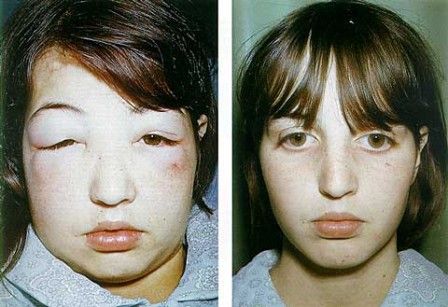
# Edema: due to decreased osmotic Pressure of blood ---> increased salt and water retention.
# Hypoalbuminemia = hyperproteinurea.


*******************************************************


Cause of Nephrotic Syndrome

Prevalence (%)[*]
Causes
Children
Adults
PRIMARY GLOMERULAR DISEASE
Membranous glomerulopathy
5
30
Minimal-change disease
65
10
Focal segmental glomerulosclerosis
10
35
Membranoproliferative
10
10
glomerulonephritis[†]
10
15
Other proliferative glomerulonephritides (focal, “pure mesangial,” IgA nephropathy)[†]
SYSTEMIC DISEASES


Amyloidosis

Systemic lupus erythematosus

Drugs (nonsteroidal anti-inflammatory, penicillamine, “street heroin”)

Infections (malaria, syphilis, hepatitis B and C, HIV)

Malignant disease (carcinoma, lymphoma)

Miscellaneous (bee-sting allergy, hereditary nephritis)


#1 Minimal-change ( glomerulnephritis ) disease:

effacement of foot processes
Causes: # follows respiratoty infection # Hodgkin lymphoma # rotine immunization # atopic disorders. بالحساسية

Pathogenesis: immune dysfunction ---> release of cytokines ----> effacement of foot ( minor ) processes ---> increased leakness.

Morpholgy: # no chanes detected by LM and immunofluorescence.
# by EM: effacement of foot processes = minimal change.

Outcomes: # no serious outcomes
# response effectively to corticosteroids.


Minimal change disease. A, Under the light microscope the PAS-stained glomerulus appears normal, with a delicate basement membrane. B, Schematic diagram illustrating diffuse effacement of foot processes of podocytes with no immune deposits.


#2 Membranous Glomerunephritis ( nephropathy ) :
chronic immune-complex mediated disease
Causes: # ideopathic ( 85 % ) 
# Autoimmune diseases ( LUPUS )
# Infections ( Malaria & Hepatitis B )
# Drugs ( NSAIDS )
# Tumours ( lung and colon )

Pathogenesis: it's chronic immune-complex mediated disease ---> deposition of this comlexes in the GBM ---> activiation of C5b-9 complement then foming membrane attack complex ---> epithelial and mesangial cells release oxidase and protease ---> attack glomerular walls ---> increase leakness of this walls. ( خناقة )


Membranous nephropathy. A, Silver methenamine stain. Note the marked diffuse thickening of the capillary walls without an increase in the number of cells. There are prominent “spikes” of silver-staining matrix (arrow) projecting from the basement membrane lamina densa toward the urinary space, which separate and surround deposited immune complexes that lack affinity for the silver stain. B, Electron micrograph showing electron-dense deposits (arrow) along the epithelial side of the basement membrane (B). Note the effacement of foot processes overlying deposits. CL, capillary lumen; End, endothelium; Ep, epithelium. C, Characteristic granular immunofluorescent deposits of IgG along GBM. D, Diagrammatic representation of membranous nephropathy

Morpholgy: Diffuse thickening of the glomerular walls = membranous.
# with LM: GBM is thickened. With advanced disease, glomeruli may be sclerosed.
# with EM: there is subepithelial GBM deposits incoporated with GBM. Effacement of foot processes.
# with immunofluorescece: diffuse granular staining of IgG and complements.
#  there is no changes in number of cells.


mmune complexes (black) are deposited in a thickened basement membrane creating a "spike and dome" appearance on electron microscopy.

Outcomes: # the patient is prsenented with NS and hematuria.
# only 10% will progress to renal failure or die.


 #3 Focal segmental glomerulosclerosis:

It characterized by involvement of some glomeruli not all ( thus, it’s focal ) and involvement of a portion of the glomerulus tuft ( so, it’s segmental ).

Causes: # Primary (idiopathic)
# secondary to known disorder ( HIV infection & Heroin addiction , obesity )
# secondary to glomerular necrosis due to other cause.
# secondary to loss of renal tissue, ( reflux nephropathy ---> urine flows backwards to kidney )
# secondary to mutations of proteins that maintain the GFB ( nephrin )

Pathogenesis: # characterized by damage of the visceral layer  ( detachment and effacement ).  
# Injury to podocytes is thought to represent the initial event that lead to release of cytokines.
# Sclerosis and hyalinosis from hyaline masses and deposition of mesangial matrix and lipids leading to obliterated capillary lumen. 
# Effacement of foot processes. It's thought that FSGS is a progression of MCD.


High-power view of focal and segmental glomerulosclerosis (PAS stain), seen as a mass of scarred, obliterated capillary lumens with accumulations of matrix material, that has replaced a portion of the glomerulus.

Collapse Glomerulopathy: # characterized by collapse of the entire glomerulus with hypertrophy of podocytes.

Collapsing glomerulopathy. Visible are retraction of the glomerular tuft, narrowing of capillary lumens, proliferation and swelling of visceral epithelial cells, and prominent accumulation of intracellular protein absorption droplets in the visceral epithelial cells


Outcomes: # the patient is prsenented with NS ,hematuria and hypertension.

# 20% will progress to renal failure or die.
#response poorly to steroids.


#4 Membranoproliferative glomerulonephritis:

Causes: # Ideopathic
# secondary to other disorder.

Types: 
# MPGN Type I: # characterized by deposition of Ab-Ag complex in the GBM # activiation of compelement system # originate from # Autoimmune diseases ( LUPUS )# Infections (  Hepatitis B )# Drugs ( NSAIDS )# Tumours ( lung and colon )
# MPGN Type II: #characterized by severe activiation of compelement system - C3 - and formation of Ab against it.


Membranoproliferative GN, 
# showing mesangial cell proliferation, 
# basement membrane thickening,
# mesangial interposition into the GBM, giving appearane of tram-track like.
# leukocyte infiltration, and accentuation of lobular architecture.
B, Schematic representation of patterns in the two types of membranoproliferative GN. 
# In type I there are subendothelial deposits;
# type II is characterized by intramembranous dense deposits (dense-deposit disease). 


Outcomes: # the patient is prsenented with NS ,hematuria and hypertension.

# 50% will progress to renal failure or die.


_______________________________________________

# Focal: some glomeruli are involved.

# Diffuse: all glomeruli are involved.

# Segmental: portion of the glomerulus is involved.

# Global: all the glomerulus is involved.


Histology of Kidney


Functional Units 
of the Kidney

# Histologicaly, kidney consists of:
 * Cortex: the outer part # granular cuz of glomeruli.
 * Medulla : the inner part # striated cuz of tubules.
 * Kidney lobule ----> papillae ----> minor calyx ----> major   calyx ----> renal pelvis ----> ureter.


Stroma :
1- Capsule
2- Reticular fibers
3- Collagenous and elastic fibers
Parynchema: uroniferous tubules:
1- nephron
2-collecting tubule


The nephron cosists of:
1- Renal corpuscle: # glomerulus # Bowman's capsule.
2- proximal tubule
3- loop of Henle's
4- distal tubule

# Nephron is functional unit of the kidney.


Diagram of renal corpuscle structure:
A – Renal corpuscle
B – Proximal tubule
C – Distal convoluted tubule
D – Juxtaglomerular apparatus
1. Basement membrane (Basal lamina)
2. Bowman's capsule – parietal layer
3. Bowman's capsule – visceral layer
3a. Pedicels (Foot processes from podocytes)
3b. Podocyte
4. Bowman's space (urinary space)
5a. Mesangium – Intraglomerular cell
5b. Mesangium – Extraglomerular cell
6. Granular cells (Juxtaglomerular cells)
7. Macula densa
8. Myocytes (smooth muscle)
9. Afferent arteriole
10. Glomerulus Capillaries
11. Efferent arteriole


# Bowman's Capsule: consists of 2 layers:
1- Parietal layer: simble squamous epi. 2
2- Visceral layer: Podocytes 3
# Mesangial cells : # macrophages # supporting cells # part of juxtaglomerular apparatus.
# Proximal tubules : # cubical # wider in diameter # narrower lumen # cells with microvilli # section shows 3-5 cells # cells are indistinct.
# Distal tubules : # cubical # wider in lumen # narrower diameter # cells without microvilli # section shows 5-8 cells # cells are distinct.
# Juxtaglomerular apparatus : # consists of:
1- JG cells: modified SM cells of afferent arteriole.
2- Macula densa: modified cells of distal tubule.
3- Polar cushion: extrglomerular mesangeal cells.
# function of JGA : secretion of Renin
Angiotensinogen ---> angiotensin I ---> angiotensin II


 Glomerular Filtration Barrier:
1- Capillary endothelium: fenestrated epi.                           
2- Fused basement membrane: of 3 layers, the middle is dark               
3- Fitration slits diaphragm: between minor - foot - processes of podocytes # covered by diaphragm.  


Nephron structure in details: