Overview of
Nephrotic Syndrome
It's increased permeability of the glomerular membrane to proteins leading to proteinuria > 3.5 g/day
Outcomes: # the patient is prsenented with NS ,hematuria and hypertension.
# 20% will progress to renal failure or die.
#response poorly to steroids.
Types:
# MPGN Type I: # characterized by deposition of Ab-Ag complex in the GBM # activiation of compelement system # originate from # Autoimmune diseases ( LUPUS )# Infections ( Hepatitis B )# Drugs ( NSAIDS )# Tumours ( lung and colon )
# MPGN Type II: #characterized by severe activiation of compelement system - C3 - and formation of Ab against it.
Membranoproliferative GN,
# showing mesangial cell proliferation,
# basement membrane thickening,
# mesangial interposition into the GBM, giving appearane of tram-track like.
# leukocyte infiltration, and accentuation of lobular architecture.
B, Schematic representation of patterns in the two types of membranoproliferative GN.
# In type I there are subendothelial deposits;
# type II is characterized by intramembranous dense deposits (dense-deposit disease).
Outcomes: # the patient is prsenented with NS ,hematuria and hypertension.
# 50% will progress to renal failure or die.
_______________________________________________
Nephrotic Syndrome
It's increased permeability of the glomerular membrane to proteins leading to proteinuria > 3.5 g/day
Symptoms:
# Hyperproteinuria : increased protien in urine > 3.5 g/day.
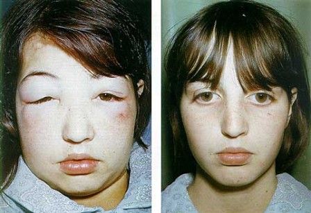
# Hyperlipidemia : incresed lipids in blood due to depletion of lipoproteins.
# Edema: due to decreased osmotic Pressure of blood ---> increased salt and water retention.
# Hypoalbuminemia = hyperproteinurea.
# Hyperlipidemia : incresed lipids in blood due to depletion of lipoproteins.
# Edema: due to decreased osmotic Pressure of blood ---> increased salt and water retention.
# Hypoalbuminemia = hyperproteinurea.
*******************************************************
Cause
of Nephrotic Syndrome
Prevalence
(%)[*]
|
|||||||||||||||||||||||
Causes
|
Children
|
Adults
|
|||||||||||||||||||||
PRIMARY GLOMERULAR DISEASE
|
|||||||||||||||||||||||
Membranous glomerulopathy
|
5
|
30
|
|||||||||||||||||||||
Minimal-change disease
|
65
|
10
|
|||||||||||||||||||||
Focal segmental
glomerulosclerosis
|
10
|
35
|
|||||||||||||||||||||
Membranoproliferative
|
10
|
10
|
|||||||||||||||||||||
glomerulonephritis[†]
|
10
|
15
|
|||||||||||||||||||||
Other proliferative
glomerulonephritides (focal, “pure mesangial,” IgA nephropathy)[†]
|
|||||||||||||||||||||||
SYSTEMIC DISEASES
|
|||||||||||||||||||||||
|
|||||||||||||||||||||||
#1 Minimal-change ( glomerulnephritis ) disease:
effacement of foot processes
Causes: # follows respiratoty infection # Hodgkin lymphoma # rotine immunization # atopic disorders. بالحساسية
Pathogenesis: immune dysfunction ---> release of cytokines ----> effacement of foot ( minor ) processes ---> increased leakness.
Morpholgy: # no chanes detected by LM and immunofluorescence.
# by EM: effacement of foot processes = minimal change.
Outcomes: # no serious outcomes
# response effectively to corticosteroids.
Minimal change disease. A, Under the light microscope the PAS-stained
glomerulus appears normal, with a delicate basement membrane. B,
Schematic diagram illustrating diffuse effacement of foot processes of podocytes
with no immune deposits.
#2 Membranous Glomerunephritis ( nephropathy ) :
chronic immune-complex mediated disease
Causes: # ideopathic ( 85 % )
# Autoimmune diseases ( LUPUS )
# Infections ( Malaria & Hepatitis B )
# Drugs ( NSAIDS )
# Tumours ( lung and colon )
Pathogenesis: it's chronic immune-complex mediated disease ---> deposition of this comlexes in the GBM ---> activiation of C5b-9 complement then foming membrane attack complex ---> epithelial and mesangial cells release oxidase and protease ---> attack glomerular walls ---> increase leakness of this walls. ( خناقة )
Membranous nephropathy. A, Silver methenamine
stain. Note the marked diffuse thickening of the capillary walls without an
increase in the number of cells. There are prominent “spikes” of silver-staining
matrix (arrow) projecting from the basement membrane lamina densa toward
the urinary space, which separate and surround deposited immune complexes that
lack affinity for the silver stain. B, Electron micrograph showing
electron-dense deposits (arrow) along the epithelial side of the basement
membrane (B). Note the effacement of foot processes overlying deposits. CL,
capillary lumen; End, endothelium; Ep, epithelium. C, Characteristic
granular immunofluorescent deposits of IgG along GBM. D, Diagrammatic
representation of membranous nephropathy
Morpholgy: Diffuse thickening of the glomerular walls = membranous.
# with LM: GBM is thickened. With advanced disease, glomeruli may be sclerosed.
# with EM: there is subepithelial GBM deposits incoporated with GBM. Effacement of foot processes.
# with immunofluorescece: diffuse granular staining of IgG and complements.
# there is no changes in number of cells.
mmune complexes (black) are deposited in a thickened basement membrane creating a "spike and dome" appearance on electron microscopy.
Outcomes: # the patient is prsenented with NS and hematuria.
# only 10% will progress to renal failure or die.
# only 10% will progress to renal failure or die.
#3 Focal segmental glomerulosclerosis:
It characterized by involvement of some glomeruli not all (
thus, it’s focal ) and involvement of a portion of the glomerulus tuft ( so,
it’s segmental ).
Causes: # Primary (idiopathic)
# secondary to known disorder ( HIV infection & Heroin
addiction , obesity )
# secondary to glomerular necrosis due to other cause.
# secondary to loss of renal tissue, ( reflux nephropathy ---> urine flows backwards to kidney )
# secondary to mutations of proteins that maintain the GFB (
nephrin )
Pathogenesis: # characterized by damage of the visceral layer ( detachment and effacement ).
# Injury to podocytes is thought to represent the initial event that lead to release of cytokines.
# Sclerosis and hyalinosis from hyaline masses and deposition of mesangial matrix and lipids leading to obliterated capillary lumen.
# Effacement of foot processes. It's thought that FSGS is a progression of MCD.
High-power
view of focal and segmental glomerulosclerosis (PAS stain), seen as a mass of scarred,
obliterated capillary lumens with accumulations of matrix material, that has
replaced a portion of the glomerulus.
Collapse Glomerulopathy: # characterized by collapse of the entire glomerulus with hypertrophy of podocytes.
Collapsing glomerulopathy. Visible are retraction of
the glomerular tuft, narrowing of capillary lumens, proliferation and swelling
of visceral epithelial cells, and prominent accumulation of intracellular
protein absorption droplets in the visceral epithelial cells
Outcomes: # the patient is prsenented with NS ,hematuria and hypertension.
# 20% will progress to renal failure or die.
#response poorly to steroids.
#4 Membranoproliferative glomerulonephritis:
Causes: # Ideopathic
# secondary to other disorder.
# secondary to other disorder.
Types:
# MPGN Type I: # characterized by deposition of Ab-Ag complex in the GBM # activiation of compelement system # originate from # Autoimmune diseases ( LUPUS )# Infections ( Hepatitis B )# Drugs ( NSAIDS )# Tumours ( lung and colon )
# MPGN Type II: #characterized by severe activiation of compelement system - C3 - and formation of Ab against it.
Membranoproliferative GN,
# showing mesangial cell proliferation,
# basement membrane thickening,
# mesangial interposition into the GBM, giving appearane of tram-track like.
# leukocyte infiltration, and accentuation of lobular architecture.
B, Schematic representation of patterns in the two types of membranoproliferative GN.
# In type I there are subendothelial deposits;
# type II is characterized by intramembranous dense deposits (dense-deposit disease).
Outcomes: # the patient is prsenented with NS ,hematuria and hypertension.
# 50% will progress to renal failure or die.
_______________________________________________
# Focal: some
glomeruli are involved.
#
Diffuse: all glomeruli are involved.
# Segmental:
portion of the glomerulus is involved.
# Global: all
the glomerulus is involved.


ليست هناك تعليقات:
إرسال تعليق